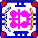
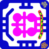
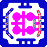
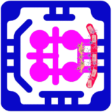
Vasculogenesis is studied by reconstructing the process through which endothelial cells self-organize into vascular networks within a biomimetic environment. These network provide nutrient and oxygen delivery, enable immune and drug transport, and connect different tissue modules. By combining endothelial cells with supporting stromal populations under physiologic flow, these platforms capture both the structural development of blood vessels and their functional integration across multiple organs.
The cellular foundation of these models typically includes human endothelial cells, pericytes, smooth muscle cells, or mesenchymal stromal cells, providing key paracrine cues to stabilize the newly formed vessels. In some applications, tissue-specific endothelial subtypes such as brain microvascular cells or liver sinusoidal endothelial cells are used to more closely reproduce organ-specific vascular physiology. The fibrin or collagen gel encapsulates the cells with angiogenic factors including VEGF, FGF-2, or angiopoietin-1. Within this microenvironment, endothelial cells extend sprouts, branch, and eventually anastomose into networks that resemble microvasculature.
The application of shear stress through perfusion flow stimulates endothelial alignment, tightens cell–cell junctions, and drives the maturation of perfusable lumens. These cues encourage pruning of unstable sprouts and stabilization of functional networks, resulting in vessels that more closely resemble in vivo physiology. The geometry of the chip provides spatial guidance, allowing vascular sprouts to connect across parenchymal and vascular compartments, thereby linking different organ modules.
Researchers assess vasculogenesis in these systems through a variety of functional readouts. Imaging reveals network morphology, branch density, and lumen diameter. Barrier integrity is measured with tracer assays such as dextran permeability, while molecular staining for markers confirms vascular identity and maturity. Perfusion studies using fluorescent beads or dyes demonstrate whether the vessels can carry flow across compartments. Importantly, these vascular networks also serve as conduits for studying immune cell trafficking, drug distribution, and metabolic cross-talk between organs.
The ability to study vasculogenesis in a multiorgan setting demonstrates that vasculogenesis is a dynamic process reproducing physiologic and pathophysiologic cross-talk in vitro. Liver–tumor platforms reveal cancer-derived VEGF alters vascular sprouting and permeability, while islet–vascular constructs highlight how microvessel density supports β-cell survival and insulin secretion. In bone marrow–vascular plates vasculogenesis provides a route for hematopoietic stem cell homing and immune cell egress, and in immuno-oncology models, it enables the study of T-cell infiltration into tumors under checkpoint blockade.


